
anti-CD52 (Mouse) Antibody, Rabbit Serum
$ 96.08
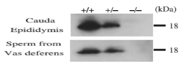
Description CD52 may play a role in carrying and orienting carbohydrate, as well as having a more specific role. Expressed on lymphohematopoietic tissues, including thymus, spleen, and bone marrow, but not in liver, kidney, and brain. Molecular mass: 7,798 Da with 74 amino acids. In mature form, propeptide is removed, and GPI anchored and glycosylated. Key words: CD52, CAMPATH-1 antigen, Lymphocyte differentiation antigen B7, Cell membrane, GPI-anchor, Glycoprotein. Specifications: Reactivity: Mouse. Likely to react with rat due to the sequence identity. Immunogen: Synthetic peptide corresponding to 29-48 amino acids of mouse CD52, C-AASGTNKNSTSTKKTPLKSG, conjugated with KLH Form: Whole rabbit antiserum added with 0.1% sodium azide. Validation: Specificity validated with KO mouse (Fig.2) Storage: Shipped at 4℃ or at -20℃. Upon arrival, spin-down and store at -20℃. Applications: Western blotting (1/1,000 dilution) Immunohistchemistry-P (1/100 dilution) Immunofluorescence staining (1/100 dilution) Database Links: uniprot/Q64389 Mouse CD52 Gene ID 23833 Mouse CD52 Reference: This antibody was described in Ref.1 and used in the following publications. Yamaguchi R et al. (2008) Cd52, known as a major maturation-associated sperm membrane antigen secreted from the epididymis, is not required for fertilization in the mouse. Genes Cells.13:851-61.WB. Fig.1 Western blotting analysis of CD52 expression in various tissues with anti-CD52 antibody. Testes, male reproductive ducts and sperm protein were extracted with lysis buffer containing Triton X-100 and subjected to western blot analysis. Western blots containing equal amounts of tissue proteins (30 µg) and sperm protein (10 µg) were reacted with anti-CD52 antibody at 1/1,000 dilution. Fig.2. Western blot analysis of CD52 in cauda epididymal lysates and sperm lysates of wild type and CD52 deficient mice. Cd52 / ( / ), Cd52 /– ( /–), and Cd52−/–(–/–). Cauda epididymis and sperm from vas deferens were lysed in lysis buffer containing 1% TritonX-100. Proteins (30 μg for cauda epididimis and 10 μg for sperm) were analyzed by western blotting with anti-CD52 antibody at 1/1,000 dilution. Fig.3. Western blot analysis of CD52 in lysates of mouse testis and sperm with anti-CD52 antibody. Proteins in the lysates (10 μg) are separated on SDS-PAGE (10-20% gradient) and electro-blotted to PVDF membrane. The membrane was reacted with anti-CD52 antibody at 1/1,000 dilution. As the second antibody, anti-rabbit IgG antibody conjugated with HRP (ab97051) was used at 1/10,000 dilution. The numbers on the right are positions of protein size markers shown in kDa. Fig.4. Immunohistochemistry of mouse testis using anti-CD52 antibody. Formalin-fixed and paraffin-embedded mouse testis Deparaffinization by LemosolRA (#122-03991, Wako, Osaka) Rehydration 100% Et-OH, 95%, 90%, 70%, DW Antigen retrieval Histo/Zyme (Cat.# k046; Diagnostic BioSystems) Washing PBST (0.25% triton X-100/PBS-) Blocking 10 % FBS / PBST 30 min 1st antibody 1/100 dilution in PBS- 4℃ O/N Washing PBS- 2nd antibody 1,000 dilution, 60 min (AF-488 goat anti-rabbit IgG (H&L) Washing PBS- 5 min, 3 times DAPI 1.0μg/mL DAPI in TBS 10 min Mount ImmunoSelect Antifading Mounting Medium (SCR-38447; Dianova) Fig. 5 Immunofluorescence staining of CD52 in NIH3T3 cells with anti-CD52 antibody. The cells were fixed in 4% paraaformaldehyde overnight. Permeabilization in 0.25% Triton X-100/PBS for 10 min Blocking in 1.5% BSA/PBS for 30 min 1st antibodies diluted 1/100 by blocking buffer and incubated over night 2nd antibody,goat, anti-mouse IgG conjugated withAlex 488 (1/1000 dilution). Nuclei were stained with DAPI.
 Anti-Calreticulin-3 / CALR3 / Calsperin Antibody, Rabbit Polyclonal
 Anti-Cdc22 / RMM1 (S. pombe) Antibody, Rabbit Polyclonal